Your Animal and plant cells under microscope images are available in this site. Animal and plant cells under microscope are a topic that is being searched for and liked by netizens today. You can Get the Animal and plant cells under microscope files here. Download all free photos.
If you’re searching for animal and plant cells under microscope images information related to the animal and plant cells under microscope topic, you have pay a visit to the ideal blog. Our website always provides you with suggestions for viewing the highest quality video and picture content, please kindly search and locate more informative video articles and graphics that fit your interests.
Animal And Plant Cells Under Microscope. Gently roll and rub the toothpick onto the top of a glass slide in an area that will be visible through the microscope. Cell is a tiny structure and functional unit of a living organism containing various parts known as organelles. Observe the cheek cells under both low and high power of your microscope. Rinse off the stain and allow the slide to dry.
 Relationships Between Mitosis in Eukaryotic Cells and From sciencing.com
Find out more with bitesize. That’s the major difference between plant and animal cells under microscope. However, the internal structure and organelles are more or less similar. Here�s a photo of a plant cell under an electron microscope. Looking at animal and plant cells under a microscope. The cytoplasm is also lightly stained containing a darkly stained nucleus at the periphery of the cell.may 26, 2021.
What are two similarities and two differences between plant and animal cells that can be seen under a microscope?
An animal cell also contains a cell membrane to keep all the organelles and cytoplasm contained, but it lacks a cell wall. Be sure to label the chloroplasts, the cell membrane, and the cell wall. Objective, introduction, procedure, result and questions will make a great experience to your students.contents:practice lab # 1: And their subsequent growth in a favorable. Put a couple of drops of methylene blue stain on the smear and leave for a few mintues. Onion epidermis under light microscope purple colored large cells project microscopic photography epidermis.
 Source: microspedia.blogspot.com
The plant cells contain chloroplast since they undergo photosynthesis but animal cells do not have this. See how a generalized structure of an animal cell. And their subsequent growth in a favorable. (ii) presence of large central vacuole in plant cell. Which characteristic can be used to tell a plant cell from an animal cell under the microscope?
 Source: brainly.in
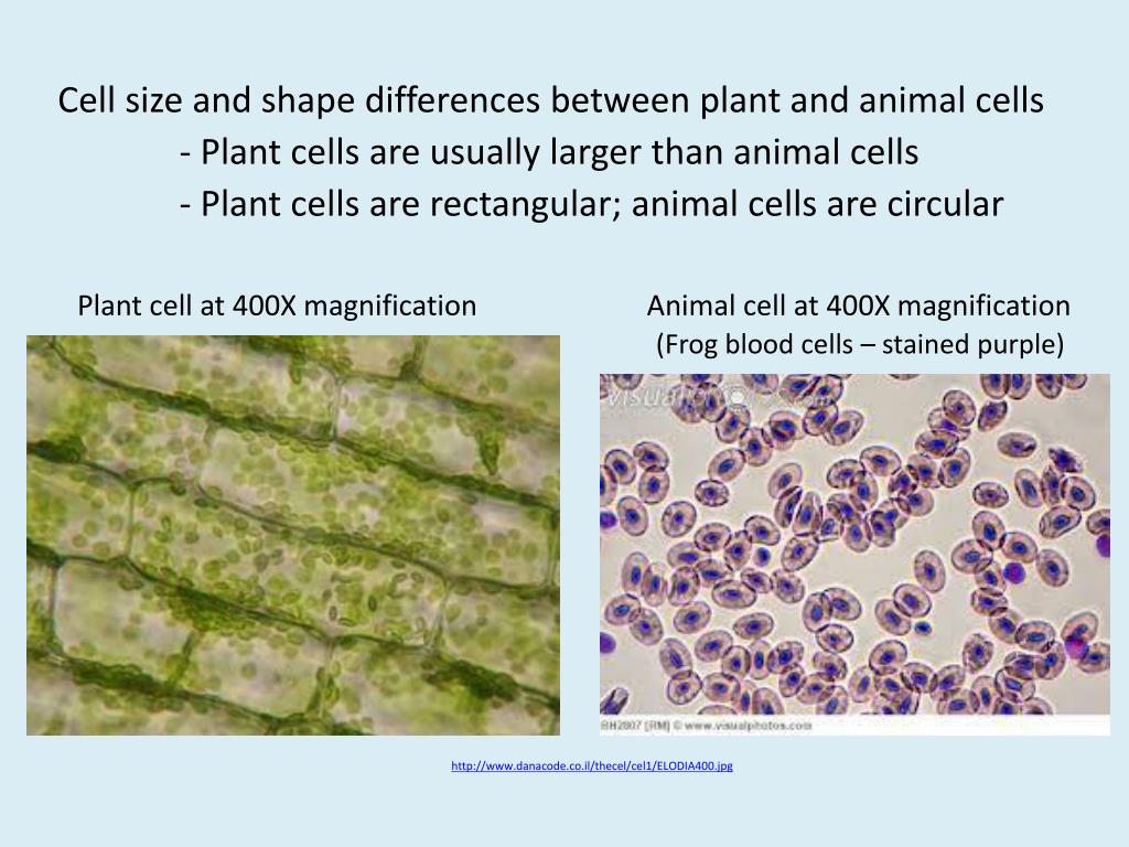
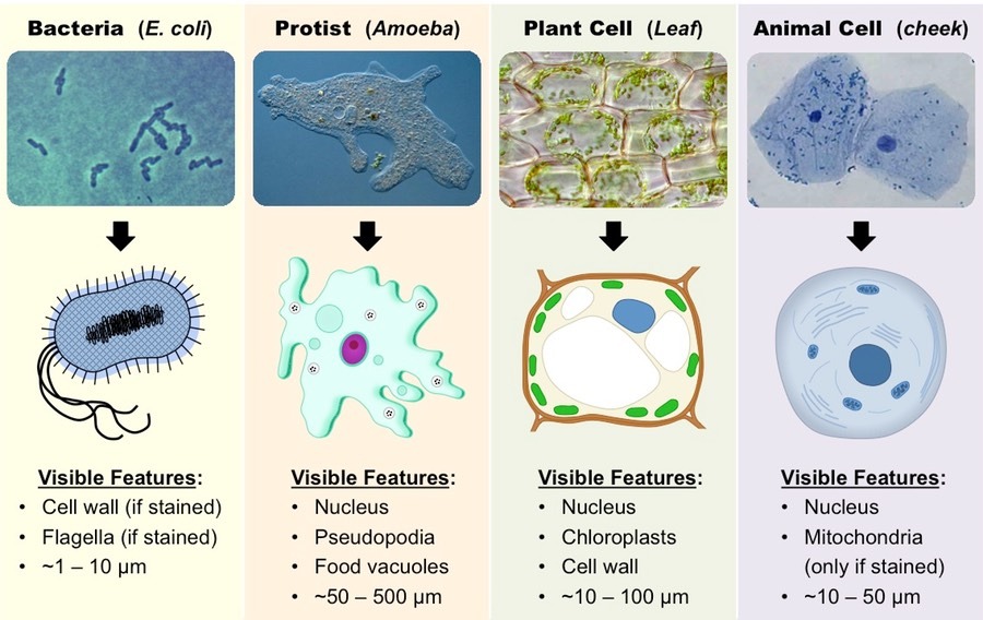
The cell also appears green in color due to the chlorophyll pigment within the chloroplasts. Under the microscope, plant cells are seen as large rectangular interlocking blocks. Cell is a tiny structure and functional unit of a living organism containing various parts known as organelles. Other organelles may also be seen, depending on the type of plant. See below to explore more:
 Source: slideserve.com
Onion epidermis under light microscope purple colored large cells project microscopic photography epidermis. Which characteristic can be used to tell a plant cell from an animal cell under the microscope? Put a couple of drops of methylene blue stain on the smear and leave for a few mintues. (ii) presence of large central vacuole in plant cell. Beneath a plant cell’s cell wall is a cell membrane.
Source: quora.com
However, the internal structure and organelles are more or less similar. What do plant and animal cells look like under a microscope? The cell also appears green in color due to the chlorophyll pigment within the chloroplasts. Here�s a photo of a plant cell under an electron microscope. For students between the ages of 11 and 14.
 Source: biologywise.com
The cell wall is somewhat thick and is seen rightly when stained. And their subsequent growth in a favorable. You will be observing plant and animal cells under the microscope. That’s the major difference between plant and animal cells under microscope. When seen under a microscope, a plant cell is somewhat rectangular in shape and displays a double membrane which is more rigid than that of an animal cell.
 Source: pinterest.com
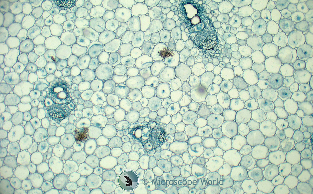
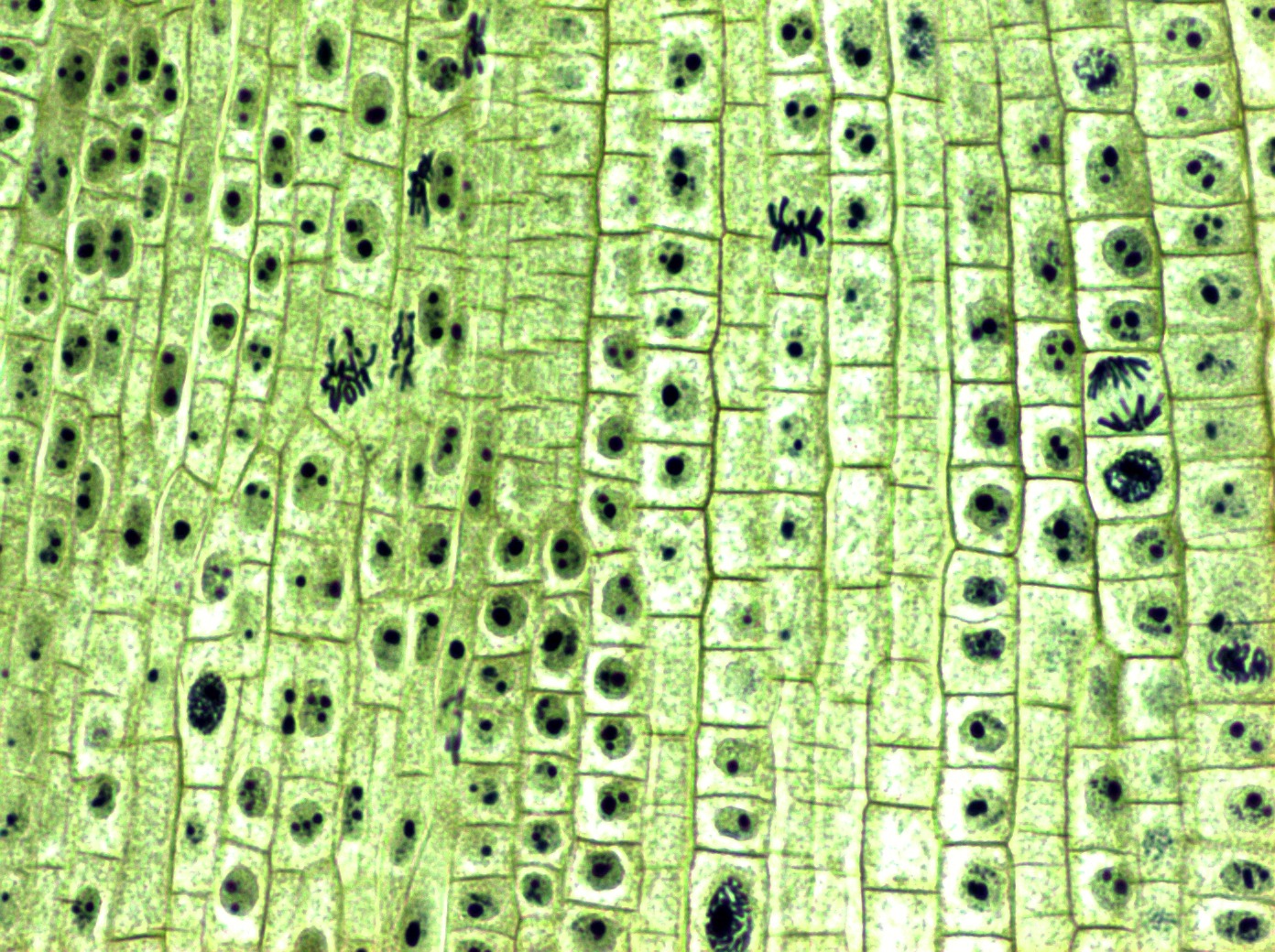
Thats the major difference between plant and animal cells under microscope. Under a microscope, plant cells from the same source will have a uniform size and shape. When seen under a microscope, a plant cell is somewhat rectangular in shape and displays a double membrane which is more rigid than that of an animal cell. What is the difference between plant and animal cells under a microscope? Gently roll and rub the toothpick onto the top of a glass slide in an area that will be visible through the microscope.
 Source: nielsonschool.blogspot.com
Thats the major difference between plant and animal cells under microscope. Discussion the focus of this practical involved making use of epithial tissue in animals and surface tissue in plants. The cell also appears green in color due to the chlorophyll pigment within the chloroplasts. Beneath a plant cell’s cell wall is a cell membrane. You can easily find samples of animal and plant cells to look at under a microscope.
 Source: pinterest.com
Gently roll and rub the toothpick onto the top of a glass slide in an area that will be visible through the microscope. Under a microscope, plant cells from the same source will have a uniform size and shape. An animal cell also contains a cell membrane to keep all the organelles and cytoplasm contained but it lacks a cell wall. The structure that can be observed under the light microscope is. The cell also appears green in color due to the chlorophyll pigment within the chloroplasts.
 Source: pinterest.com
You can easily find samples of animal and plant cells to look at under a microscope. And their subsequent growth in a favorable. (iii) presence of cell wall. View the leaf under low, medium, and high power objectives, and then draw the cells in figure 2.2, along with any organelles you can see. The students noticed the differences and similarities between the animal and plant cell under microscope.
 Source: pulpbits.net
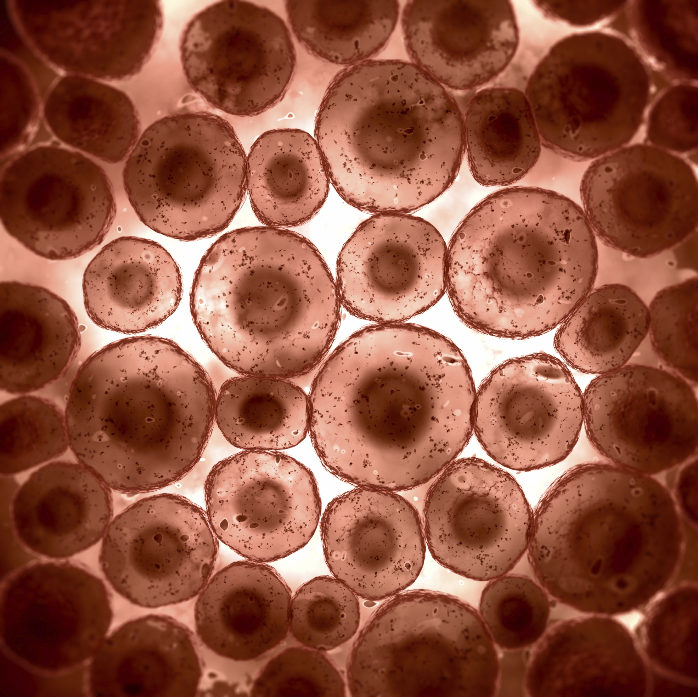
Under the microscope animal cells appear different based on the type of the cell. It was not until good light microscopes became available in the early part of the nineteenth century that all plant and animal tissues were discovered to be aggregates of individual cells. You know, animal cell structure contains only 11 parts out of the 13 parts you saw in the plant cell diagram, because chloroplast and cell wall are available only in a plant cell. Within the cell, there is a shape of round with a circular structure of granulated part on the epithelial cells. An animal cell also contains a cell membrane to keep all the organelles and cytoplasm contained, but it lacks a cell wall.
 Source: clairciccarelloe03349.blogspot.com
Cell is a tiny structure and functional unit of a living organism containing various parts known as organelles. It was not until good light microscopes became available in the early part of the nineteenth century that all plant and animal tissues were discovered to be aggregates of individual cells. An animal cell also contains a cell membrane to keep all the organelles and cytoplasm contained, but it lacks a cell wall. Be sure to label the chloroplasts, the cell membrane, and the cell wall. Beneath a plant cell’s cell wall is a cell membrane.
 Source: microspedia.blogspot.com
To prepare animal cells for viewing under a microscope. (iii) presence of cell wall. The granulated area is the cell cytoplasm while the huge round part is the nucleus. However , plant and animal cells do not exactly look the same or do not. An animal cell also contains a cell membrane to keep all the organelles and cytoplasm contained, but it lacks a cell wall.
 Source: sanimale.blogspot.com
An animal cell also contains a cell membrane to keep all the organelles and cytoplasm contained but it lacks a cell wall. Beneath a plant cell’s cell wall is a cell membrane. (iii) presence of cell wall. The cell also appears green in color due to the chlorophyll pigment within the chloroplasts. Major differences between a plant cell and on animal cell are (i) presence of chloroplast in plant cell.
 Source: solidsmack.com
Structurally, plant and anmimal cells are very similar because they are both eukaryotic cells. To prepare animal cells for viewing under a microscope. Here�s a photo of a plant cell under an electron microscope. How can you identify an animal cell under a. The cell wall is somewhat thick and is seen rightly when stained.
 Source: pinterest.co.uk
See how a generalized structure of an animal cell. To prepare animal cells for viewing under a microscope. Add a drop of purple stain (specific for animals) and cover with a cover slip. How can you identify an animal cell under a. The plant cells contain chloroplast since they undergo photosynthesis but animal cells do not have this.
 Source: sciencing.com
An animal cell also contains a cell membrane to keep all the organelles and cytoplasm contained, but it lacks a cell wall. When you look at an animal or plant cell under a microscope the most obvious feature you will see is the large dark nucleus. An animal cell also contains a cell membrane to keep all the organelles and cytoplasm contained, but it lacks a cell wall. Be sure to label the chloroplasts, the cell membrane, and the cell wall. Use a cotton swab to get cheek cells and smear this onto a slide.
 Source: pulpbits.net
It was not until good light microscopes became available in the early part of the nineteenth century that all plant and animal tissues were discovered to be aggregates of individual cells. The cell also appears green in color due to the chlorophyll pigment within the chloroplasts. Under the microscope, animal cells appear different based on the type of the cell. Plant and animal cells can be seen with a microscope. Here�s a photo of a plant cell under an electron microscope.
 Source: ttlbiology.blogspot.com
You can observe this epithelial animal cell under microscope with high power. Within the cell, there is a shape of round with a circular structure of granulated part on the epithelial cells. An animal cell also contains a cell membrane to keep all the organelles and cytoplasm contained but it lacks a cell wall. All you need to do is to gently scrape the inside of the mouth using a clean, sterile. Plant and animal cells can be seen with a microscope.
This site is an open community for users to share their favorite wallpapers on the internet, all images or pictures in this website are for personal wallpaper use only, it is stricly prohibited to use this wallpaper for commercial purposes, if you are the author and find this image is shared without your permission, please kindly raise a DMCA report to Us.
If you find this site helpful, please support us by sharing this posts to your favorite social media accounts like Facebook, Instagram and so on or you can also save this blog page with the title animal and plant cells under microscope by using Ctrl + D for devices a laptop with a Windows operating system or Command + D for laptops with an Apple operating system. If you use a smartphone, you can also use the drawer menu of the browser you are using. Whether it’s a Windows, Mac, iOS or Android operating system, you will still be able to bookmark this website.


